-
Khanna Traders, are producer and supplier of scientific equipment.
- khannachahat05@gmail.com
-
+91-8930598097


Khanna Traders, are producer and supplier of scientific equipment.
+91-8930598097
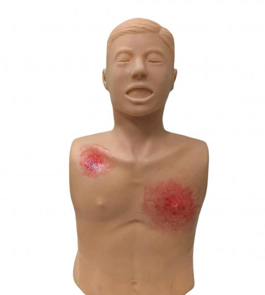
The Bandaging Training Manikin is a realistic upper-torso simulator designed for teaching wound dressing, trauma care, and bandaging techniques. The model features simulated wounds on the chest and shoulder regions, enabling nursing, medical, paramedical, and first-aid students to practice dressing and bandaging procedures in a safe and controlled environment.
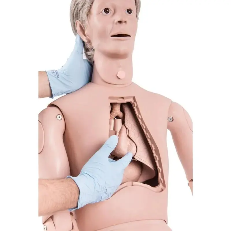
The Adult Auscultation Trainer is an advanced clinical skills simulator designed to teach and assess heart, lung, and bowel sound auscultation. The trainer provides realistic anatomical landmarks and programmable physiological sounds, enabling students and healthcare professionals to develop accurate diagnostic listening skills using a standard stethoscope.
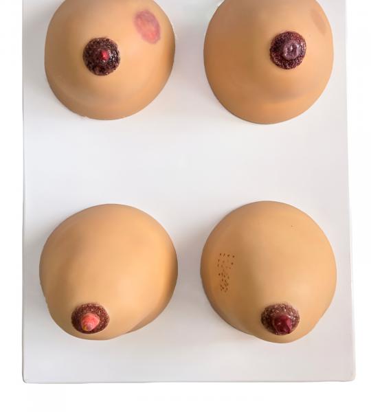
The Breast Examination Model is a realistic training simulator designed for teaching and practicing clinical breast examination (CBE), breast self-examination (BSE), and the identification of common breast abnormalities. It enables nursing, medical, and allied health students to develop essential diagnostic and palpation skills in a safe and controlled learning environment.
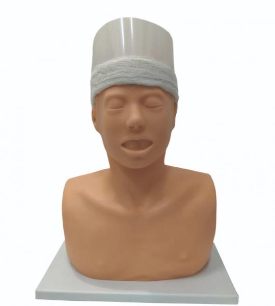
The Shiro Basti Training Manikin is a specialized Ayurveda and Panchakarma teaching aid designed for demonstrating and practicing Shiro Basti, a classical Panchakarma procedure in which medicated oil is retained on the head using a specially designed cap or reservoir. The manikin provides a realistic platform for students to learn procedural techniques safely and effectively.
Product Overview
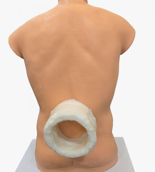
The Kati–Manya–Prushta Basti Training Manikin is designed for Ayurveda and Panchakarma practical training. It enables students to learn and practice the procedures of Kati Basti (lumbar region), Manya Basti (cervical region), and Prushta Basti (full back/spinal region) in a realistic and hygienic training environment. These therapies involve retaining warm medicated oil over specific anatomical regions using a dough ring.
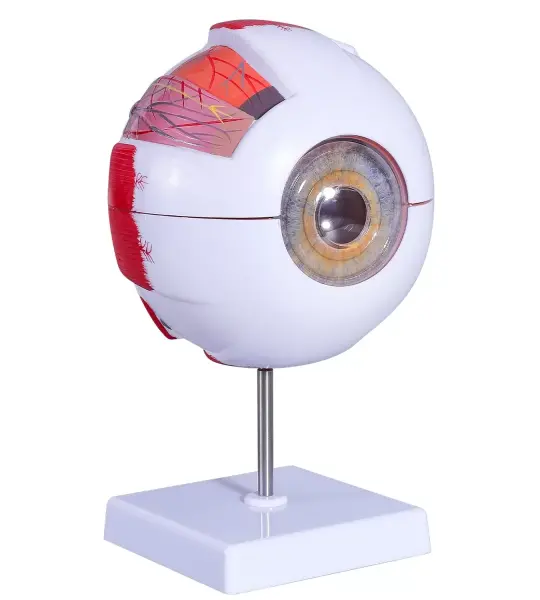
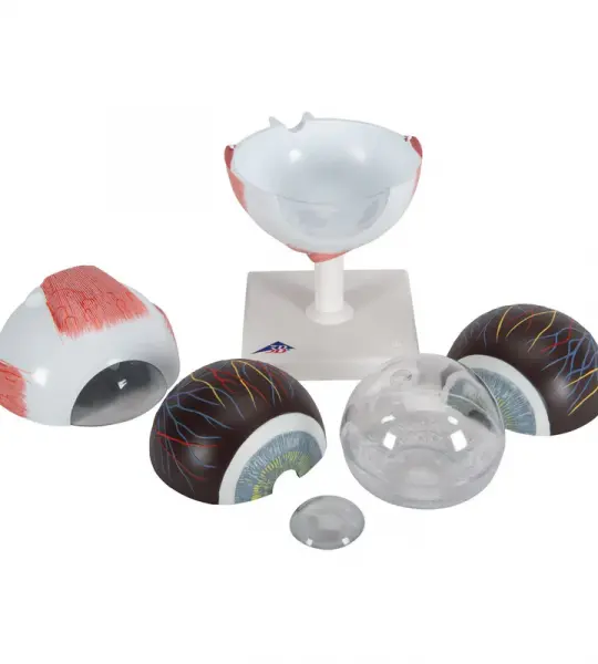
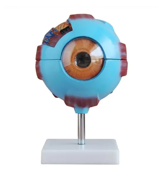
The Giant Eye replica is a great tool to teach-learn the anatomy of the eye! Removable parts of the human eye model include Lens, iris, Vitreous humour, retina
This large anatomical human eye model shows the optic nerve in its natural position in the bony orbit of the eye (floor and medial wall). At three times life size this eye model is great for anatomical demonstrations.
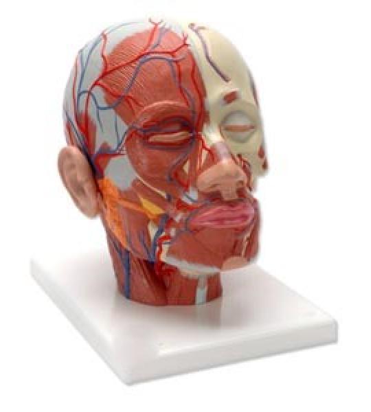
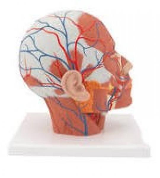
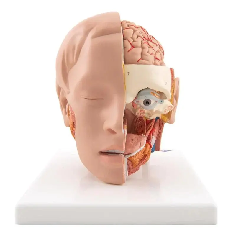
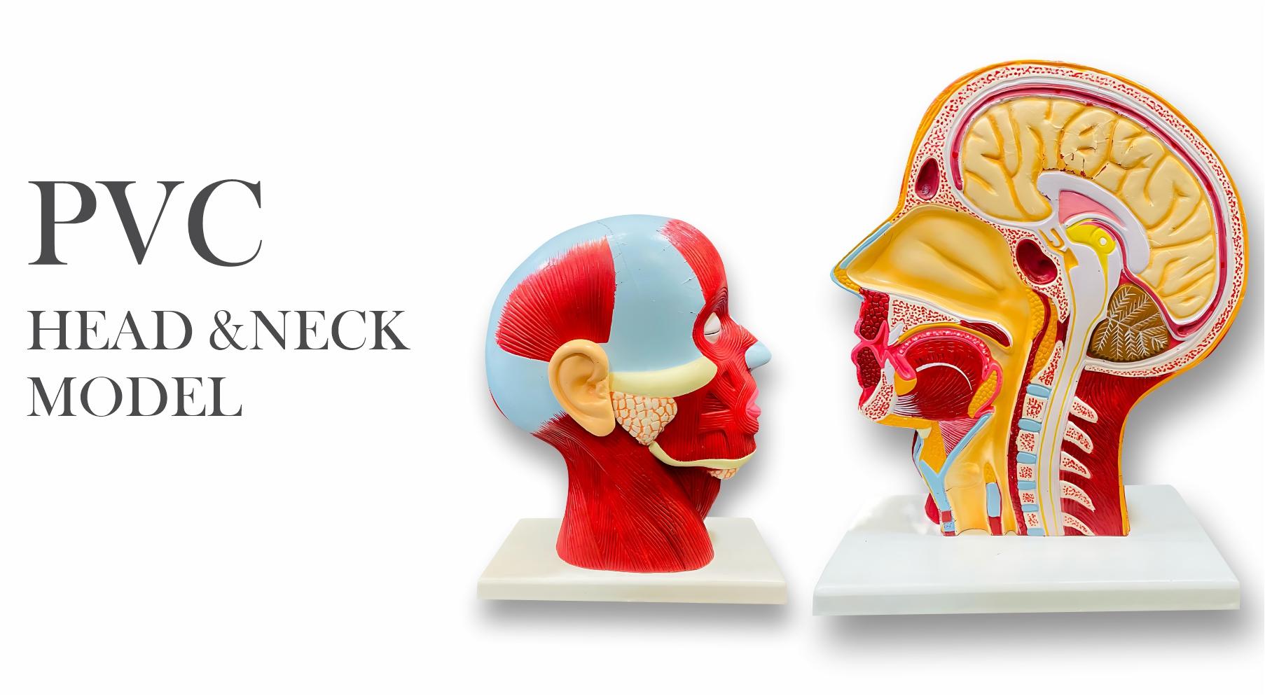
Representation of the superficial musculature with Parotid gland, Submandibular gland (right half), Deep musculature (left half).
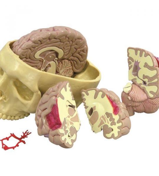
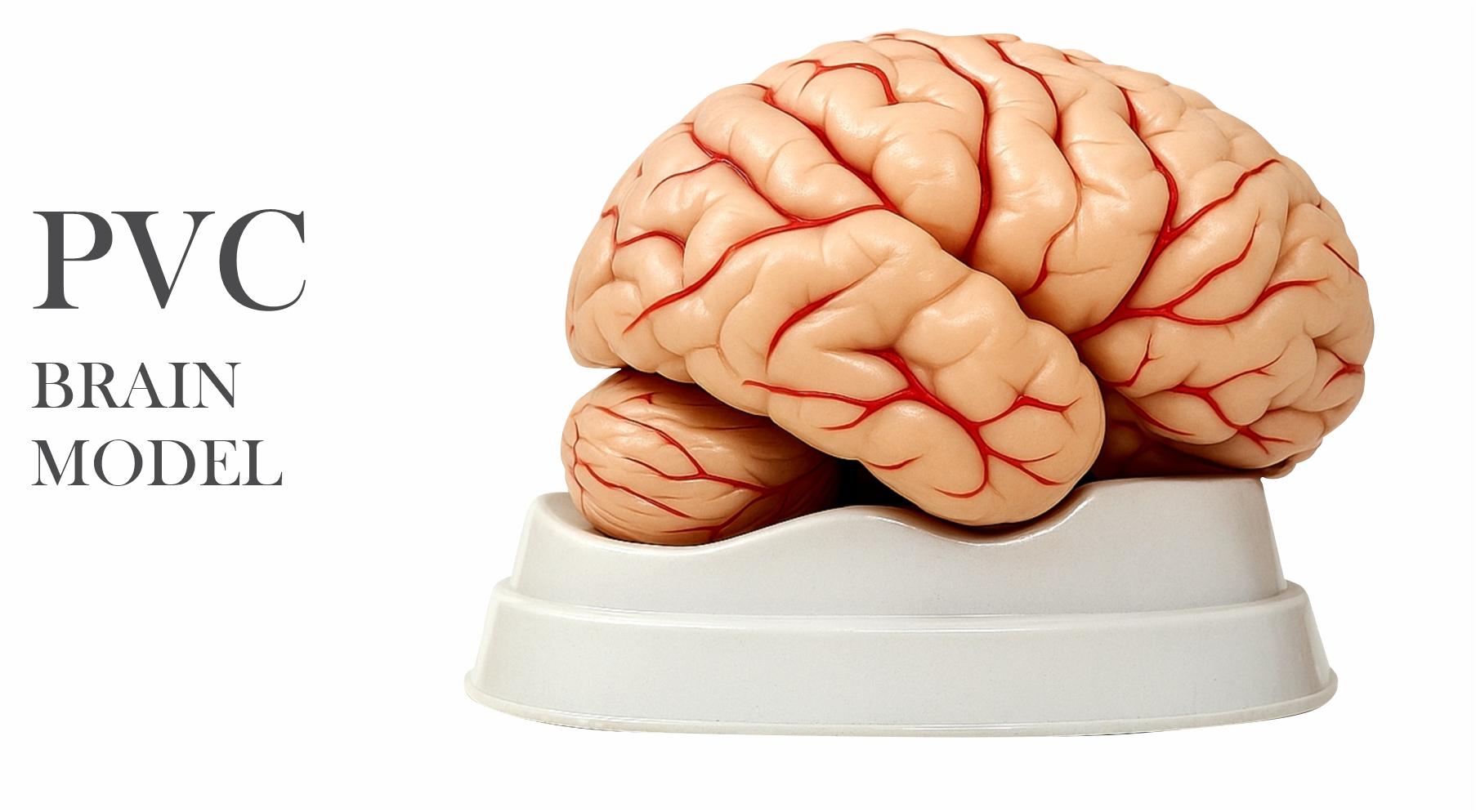
Full size segmented brain features half normal side and three-piece sectioned pathology half, as well as Circle of Willis with aneurism. The brain, which sits inside a partial skull, features the following pathologies which are also illustrated on a two-sided education card: alcoholism, Alzheimer’s, aneurism, depression related tumor, seizure related tumor, migraine, multiple sclerosis, Parkinson’s disease, stroke, and subdural hematoma.
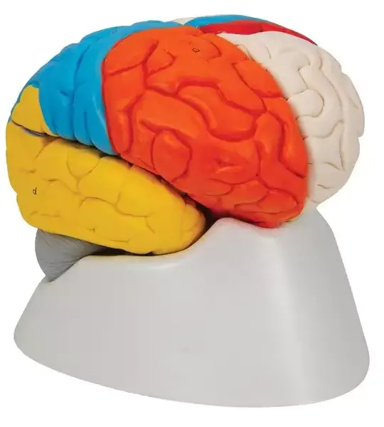
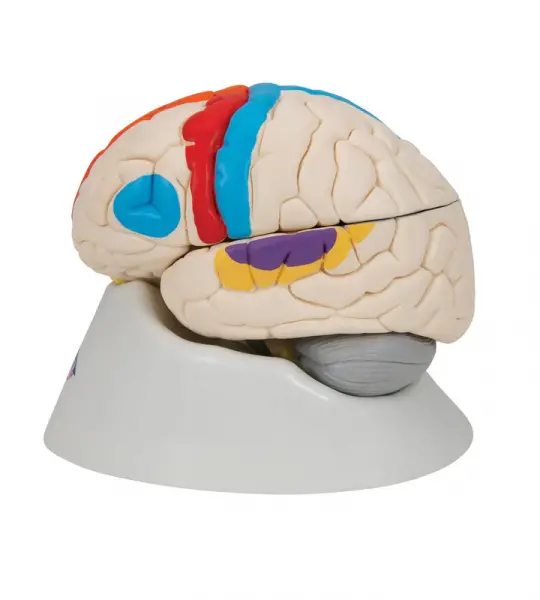
This deluxe brain is medially divided. On the right half of this brain, you will find a colored, systematic grouping and representation of the cerebral lobe.

The Giant Eye replica is a great tool to teach-learn the anatomy of the eye! Removable parts of the human eye model include Lens, iris, Vitreous humour, retina
This large anatomical human eye model shows the optic nerve in its natural position in the bony orbit of the eye (floor and medial wall). At three times life size this eye model is great for anatomical demonstrations.
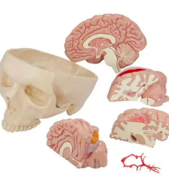
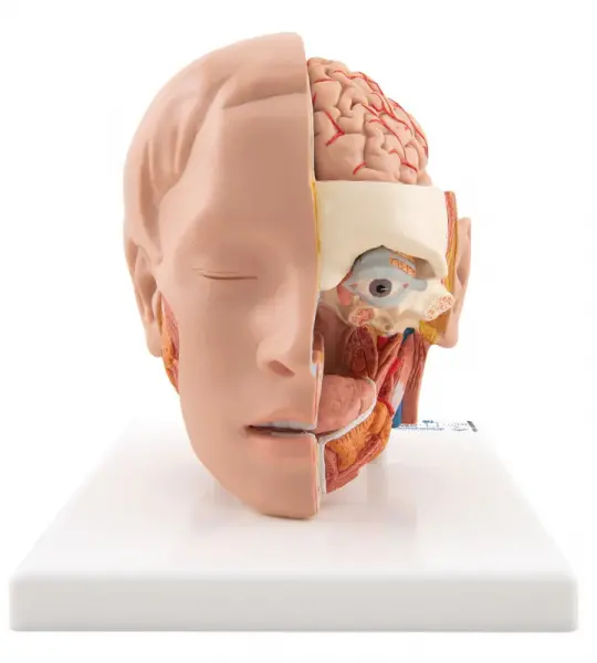
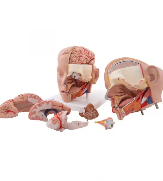
Our most detailed head model! This life-size 6-part head is mounted on a base and features a removable 4-part brain half with arteries.
Full size segmented brain features half normal side and three-piece sectioned pathology half, as well as Circle of Willis with aneurism. The brain, which sits inside a partial skull, features the following pathologies which are also illustrated on a two-sided education card: alcoholism, Alzheimer’s, aneurism, depression related tumor, seizure related tumor, migraine, multiple sclerosis, Parkinson’s disease, stroke, and subdural hematoma.
This deluxe brain is medially divided. On the right half of this brain, you will find a colored, systematic grouping and representation of the cerebral lobe.
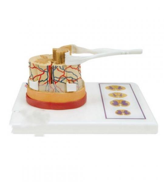
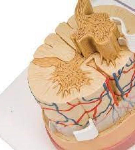
The Spinal cord model illustrates the composition of the spinal cord, magnified to a scale of about 5:1. The spinal cord is formed by a central channel surrounded by "gray matter" with an outer layer of "white matter".